Most medical imaging (X-Rays, CT scans) is like a photograph of a house: it shows you the walls, the roof, and the foundation.
But a PET Scan (Positron Emission Tomography) is different. It doesn't just show the house; it shows the lights turning on, the heater running, and the activity in the kitchen. It is a living picture of your body’s metabolism.
By using a tiny amount of radioactive tracer, a PET scan tracks where your body is consuming energy in real-time. It allows doctors to see disease (like cancer, heart disease, or brain disorders) not just as a physical lump, but as a biochemical process in motion.
In the 1970s, a team wove together successive breakthroughs to produce the first modern PET scan.
Today, over 2 million scans a year in the U.S. provide the critical data needed to inform lifesaving treatments.
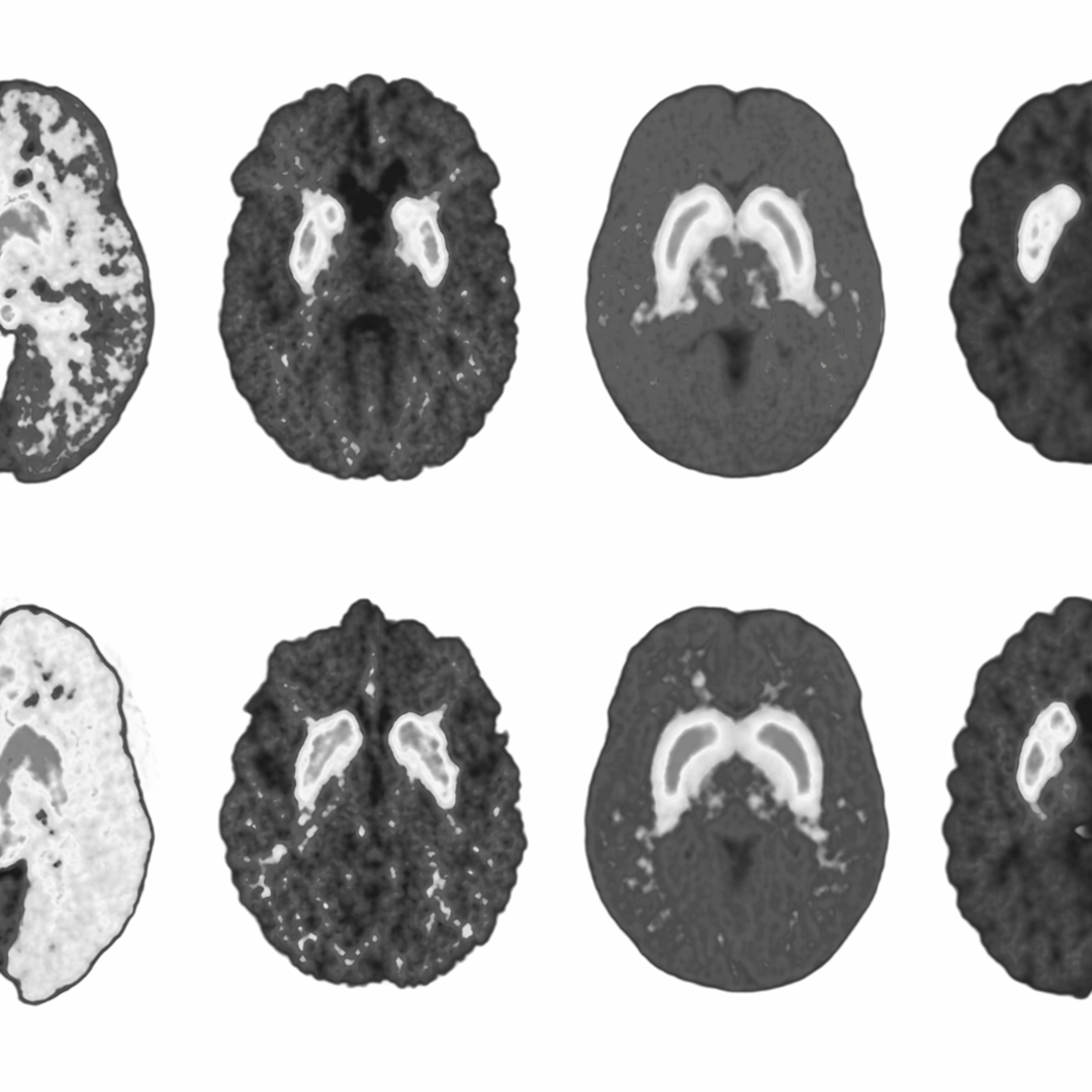
A team from St. Louis produced the first functional images of living human brain metabolism at Mass General.


Twenty-five health innovations that changed the world.



For patients and their families, a PET scan is synonymous with a moment of profound terror or suspense. It is the test you wait for when the stakes are at their highest, the one that confirms if a treatment is working or if a disease has moved.
I have lived through that suspense very recently with a health issue in my own family. Standing on that side of the glass, you don’t think about the scintillation detectors or the radiochemistry. You only think about the answer.
Having seen the clarity those images provide, I have a newfound, deep appreciation for this technology. It is because we can map metabolism in motion that we can address disease with such surgical precision. 1977 gave us the eyes to see what was once invisible, and it is saving lives, including those closest to me, every single day.